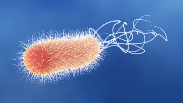
Pseudomonas aeruginosa is a Gram-negative, rod-shaped bacterium, commonly found in soil and water, and is capable of causing a wide range of infections in humans.
The Gram stain procedure is an important tool in the diagnosis of Pseudomonas aeruginosa as it helps to differentiate between Gram-positive and Gram-negative bacteria.
We will discuss the Gram stain procedure for Pseudomonas aeruginosa and its importance in the diagnosis of the bacterium. Characterized by its blue-green pigment, this bacterium is an important human and animal pathogen.
Tools for gram staining
- Glass slides
- Gram stain reagents
- Wire loop
- Water
- Burner
Pseudomonas aeruginosa gram staining procedure

1. Preparation of the bacterial smear:
A bacterial smear must first be prepared before performing a gram stain. This entails placing a small amount of the Pseudomonas aeruginosa specimen on a microscope slide.
The specimen is then spread out to form a thin, even layer of bacterial cells, known as a bacterial smear. The smear is then heat-fixed, which involves passing the slide three times through the flame of a Bunsen burner.
Heat fixing kills the bacteria, causes them to adhere to the slide, and coagulates the bacterial proteins, preventing them from being washed away during the staining process.
2. Primary Stain:
It is the stain applied to the Pseudomonas aeruginosa smear staining procedure. In this case, the primary stain is crystal violet. It is a basic dye that binds to the negatively charged bacterial cell wall.
Crystal violet is added to the Pseudomonas aeruginosa smear and allowed to sit for one minute. During that time, the stain will penetrate all bacterial cell walls, turning them purple when viewed under a light microscope.
3. Mordant:
Next, an iodine solution is applied to the smear as a mordant, which means that it helps to intensify the staining by binding the crystal violet more tightly to the bacterial cells.
Pseudomonas aeruginosa gram stain reaction: Inside the bacterial cell wall, the iodine solution forms a stable complex with crystal violet.
This helps to keep the dye from washing away during the subsequent decolorization step. The decolorization step entails washing the smear with alcohol or acetone, which removes crystal violet from some but not all bacterial cells.
4. Decolorizer:
Typically, ethanol or acetone is used as a decolorizer in the Gram staining procedure.
The decolorizer is added after the mordant and is responsible for removing the crystal violet-iodine complex from the outer membrane of Pseudomonas aeruginosa, whereas the complex remains trapped within Gram-positive bacteria’s thicker peptidoglycan layer.
The bacterial cell wall’s differential affinity for the crystal violet-iodine complex and the decolorizer is what allows the final staining color to distinguish between the two groups – Gram-positive bacteria appear purple, while Gram-negative bacteria appear pink.
5. Counterstain:
Finally, the smear is stained with a counterstain such as safranin. The safranin colorizes the decolorized Pseudomonas aeruginosa cells pink-red, whereas the crystal violet-iodine complex remains in the intact cell walls of other bacterial cells, which appear purple-blue.
This differential staining process distinguishes Gram-positive bacteria, which retain the crystal violet-iodine complex and appear purple-blue after counterstaining, from Gram-negative bacteria, which do not retain the complex and appear pink-red.
Pseudomonas aeruginosa gram stain morphology
The morphology of Pseudomonas talks about its shape or structure after gram staining was done on it. When viewed under a microscope, Pseudomonas aeruginosa appears as a rod-shaped bacterium with a single flagellum.
Pseudomonas aeruginosa gram stain cell arrangement
Depending on the growth conditions and stage of development, the arrangement of these cells may vary. Cells can be found singly or in pairs, short chains, or clusters.
Pseudomonas aeruginosa gram stain color
Pseudomonas aeruginosa will appear as reddish or pink rods under the microscope after a Gram stain, indicating that they are Gram-negative bacteria.
Gram stain picture of pseudomonas aeruginosa
Pseudomonas aeruginosa under the microscope after gram staining reveals a pink coloration.
Pseudomonas macconkey agar
To culture, Pseudomonas aeruginosa, a selective and differential medium such as MacConkey agar can be used. MacConkey agar contains crystal violet and bile salts, which inhibit the growth of Gram-positive bacteria and allow only Gram-negative bacteria to grow.
In addition, MacConkey agar contains lactose and a pH indicator that changes color when lactose is fermented. Pseudomonas aeruginosa is a lactose non-fermenter, which means it does not change the color of the agar.
Pseudomonas aeruginosa is cultured on MacConkey agar by streaking the bacteria sample onto the agar and then placing the plate in an incubator to allow the bacteria to grow.
The presence of Pseudomonas aeruginosa on agar result can be confirmed after incubation by its characteristic appearance, which is a colorless colony with no change in the color (pink) of the surrounding medium.
The following steps are involved in the culture of Pseudomonas aeruginosa on MacConkey agar:
- Prepare the MacConkey agar according to the manufacturer’s instructions and sterilize it by autoclaving.
- Using a sterile loop or swab, inoculate the agar with a sample of the specimen suspected of containing Pseudomonas aeruginosa.
- Incubate the agar for 18-24 hours at 37°C.
- After incubation, observe the agar for the growth of Pseudomonas aeruginosa. Pseudomonas aeruginosa appears as pink, flat, and circular colonies with a smooth surface on MacConkey agar.
- Biochemical tests should be performed to confirm the identification of Pseudomonas aeruginosa.
Pseudomonas aeruginosa on nutrient agar
Nutrient agar is a general-purpose growth medium that can support the growth of a variety of microorganisms, including Pseudomonas aeruginosa.
The following are the steps involved in cultivating Pseudomonas aeruginosa on nutrient agar: Follow steps 1 to step 5 as outlined for Macconkey agar.
On nutrient agar, Pseudomonas aeruginosa appears as a round, smooth, and glistening colony with a bluish-green color.
Pseudomonas aeruginosa on chocolate agar
Chocolate agar, a non-selective, enriched medium commonly used for the isolation and growth of fastidious microorganisms, can be used to culture Pseudomonas aeruginosa.
Chocolate agar is created by combining lysed red blood cells with nutrient agar, which provides fastidious microorganisms with the necessary nutrients and growth factors.
Because of the heat used in preparing the agar and lysing the red blood cells, the agar turns brown, earning it the name “chocolate agar.”
Follow steps 1 to 5 as outlined for Macconkey agar if you want to culture Pseudomonas aeruginosa on chocolate agar.
On chocolate agar, Pseudomonas aeruginosa appears as a round, smooth, and glistening colony with a greenish-blue color.
Pseudomonas aeruginosa colony
Pseudomonas aeruginosa colonies on nutrient agar appear as bluish-green colonies with a metallic sheen. The colonies appear as pink, flat, circular colonies with a smooth surface on MacConkey agar.
The colonies on blood agar are non-hemolytic and appear as small, smooth, bluish-green colonies. The colonies appear as greenish-blue colonies with smooth and shiny surfaces on chocolate agar.
What Next?
Carrying out gram staining on Pseudomonas aeruginosa is very straightforward. With the help of a lab attendant, you can get it done in a matter of minutes.
