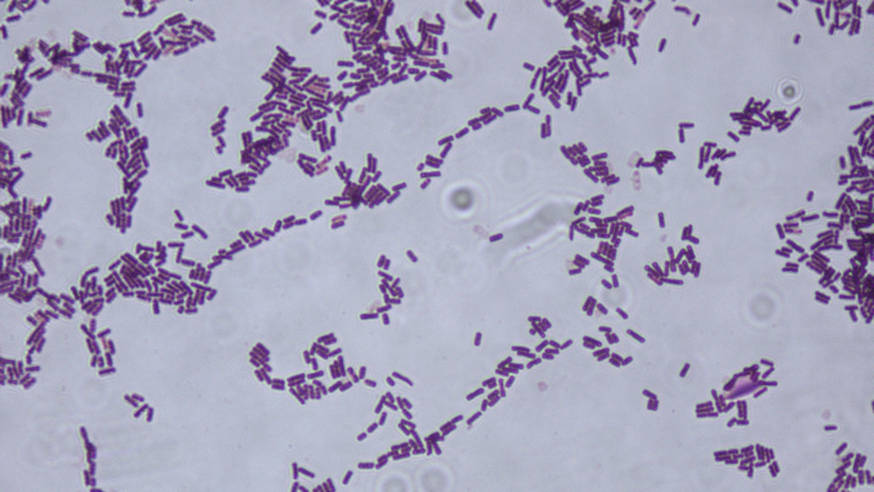
Can you use the gram staining protocol on mycoplasma?
The gram staining protocol is an important tool for identifying bacteria and other microorganisms. This article will go over how to use the Gram staining protocol on Mycoplasma, a genus of bacteria known for its small size and lack of a cell wall.
We will look at the challenges of using the Gram staining protocol on this particular genus of bacteria, as well as some potential solutions.
What is the gram staining protocol?
The gram staining protocol is also known as the gram staining procedure or method. It derives its name from Christian Gram in 1882 when he first introduced it as a way of identifying microorganisms.
It is a vital procedure used to identify and classify different types of bacteria in the field of microbiology. It is so fundamental that all microbiologists must be familiar with the procedure.
Gram stain procedure steps

Gram staining involves some steps and these steps include; the use of dyes, stains, and reagents to distinctively differentiate between gram-positive and gram-negative bacteria. These are the two major groups of bacteria in microbiology.
The gram staining procedure exploits the difference in bacteria’s cell wall thickness to identify them as gram-positive or gram-negative. Gram-positive bacteria, have thick cell walls and are more resistant to environmental stress.
On the other hand, gram-negative bacteria have a thinner cell wall which consequently makes them susceptible to environmental stress.
The gram staining procedure is as follows;
- The primary stain
- The mordant
- The decolorizer
- The counterstain
First, the bacteria sample to be identified is treated with the primary stain – crystal violet dye after a smear has been made on a slide. The smear is either air-dried or heat fixed. The aim of the crystal violet dye is for it to interact with the cell wall of the bacteria.
In the next step, the mordant iodine is added to the slide in order to fix the crystal violet dye to the cell wall of the bacteria. This interaction of iodine and the primary stain forms an insoluble complex that gets trapped in the cell wall.
In the next step, the decolorizer alcohol or acetone is added. The aim is to use it to remove the primary stain from the gram-negative bacteria but not from the gram-positive bacteria because the gram-negative bacteria have a higher lipid content than the gram-positive.
Finally, safranin (red colored), a counterstain is added to the slide. For gram-negative cells, a pink or red coloration is seen because the cells were initially decolorized but the gram-positive cells retain the purple coloration of crystal violet.
The importance of gram staining. Why is gram staining important for classifying bacteria?
The importance of the application of the gram staining technique cannot be overemphasized because, in the diagnosis and treatment of bacterial infections, it is important that the pathogenic bacteria are identified by a microbiologist before a doctor can recommend an effective treatment.
Gram staining is also important because it is a process used for the identification and classification of microorganisms. Without classification, it will be impossible for scientists to understand the roles of microorganisms in various environments.
It will also be impossible for microbiologists to identify new species and learn about their genetic makeup and evolutionary history which will make them useful in the development of new products for industrial and agricultural use.
Healthcare professionals accurately identify antibiotic-resistant bacteria using this procedure as well. Gram staining has played and is playing a fundamental role in the battle against antibiotic resistance because It can provide information about the structure of the cell wall, which can help explain how certain antibiotics can work against it.
Without gram staining, it is impossible to accurately determine the bacteria associated with a specific disease. In the instance of a disease outbreak, this procedure becomes extremely important.
Label tools used in gram staining
- A microscope
- Microscope slide
- Slide rack
- Bunsen burner
Types of gram staining
Gram staining is of two types namely;
- Simple staining
- Differential staining
The simple staining technique is an effective and straightforward method of determining the morphology, size, and arrangement of bacteria cells.
Notably, this staining technique utilizes a single dye, such as crystal violet, acridine orange, eosin, malachite green, safranin, coomassie blue, ethidium bromide, or methylene blue to color the bacterial cells and make them visible to the naked eye.
Differential staining uses more than one dye to differentiate between different types of microorganisms. Differential staining usually requires multiple steps. Gram staining is an example of differential staining than simple staining.
What is mycoplasma
This bacteria is a unique type of bacteria that is different from other bacteria because it lacks a cell wall. Mycoplasma is responsible for a number of diseases in humans including mycoplasma pneumonia, a type of bacterial pneumonia.
It is one of the smallest and simplest self-replicating organisms and it is found in soil, water, human, and animal hosts.
Can you use the gram staining protocol on mycoplasma?
We’ve successfully explored what gram staining is and also delved into what mycoplasma is. The unique aspect of mycoplasma is that it doesn’t have a cell wall howbeit, gram staining is done on bacteria with cell walls and this is used to determine if they are gram-negative or gram-positive.
Therefore the gram staining protocol is not useful in determining if the mycoplasma species are gram-negative or gram-positive because they lack a cell wall.
Studies however show that mycoplasmas evolved by degenerative evolution from Gram-positive bacteria and are phylogenetically most closely related to some clostridia.
The lack of a cell wall makes it difficult for microbiologists to use the gram staining protocol for the identification of this bacteria.
Hence other methods have been devised for the identification of this bacteria. In the case of M pneumonia, a pathogen, the fluorescent antibody technique was used for its identification.
The principle of the fluorescent test is that it uses fluorescently-labeled antibodies to detect the presence of specific antigens in a sample.
What Next?
The gram staining protocol clearly cannot be used for the identification of mycoplasma species.
FAQ
What is the theory of gram staining
The theory of gram staining involves the ability of the bacterial cell to be identified to retain the primary stain, crystal violet, within its cell wall after the primary stain has been applied.
What is the primary stain?
The primary stain is used in the first step of gram staining. The primary stain is called crystal violet.
Why do gram-negative bacteria stain red
Gram-negative bacteria stain red because the decolorizing agent, alcohol, removes the color of the primary stain it was initially treated with and then goes ahead to retain the color of the counterstain (red).
What are the four reagents used in the gram stain?
The four reagents used in the gram stain include; Crystal violet, iodine, alcohol, and safranin.
Gram stain results in interpretation
The bacterial cells that appear purple in color are gram-positive and the bacterial cells that appear pink or red are gram-negative.
Read Next
Can I study medicine after microbiology in Nigeria?
15 Benefits of studying microbiology in Nigeria
History of microbiology in Nigeria
Is microbiology a good career in Nigeria
Can you use the gram staining protocol on mycoplasma
